Chronic wounds are notoriously stubborn, and the standard playbook for treating them often relies on guesswork—but a newly published algorithm aims to replace subjective judgment with hard data. The PRISM framework, designed around Kent Imaging’s bedside imaging devices, gives clinicians a structured pathway to tackle the three silent drivers of amputation: poor blood flow, hidden bacteria, and overlooked structural problems.
Clinician-authored framework turns tissue oxygenation and bacterial autofluorescence imaging into eight point-of-care treatment scenarios.
The PRISM algorithm helps medical professionals in the fight against hard to heal ulcerations.”— Ray Abdo, DPM
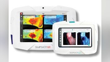
CALGARY, AB – ALBERTA, CANADA, October 29, 2026 /EINPresswire.com/ — Kent Imaging Inc., the company behind the SnapshotNIR and SnapshotGLO point-of-care wound imaging systems, announced the publication of the PRISM clinical algorithm for chronic wound care in the Journal of Wound Care.
Also worth a look: FIDER tackles tricky odd-form electronics assembly with precision pick-and-place automation and FIDER shows how to sync feeding, vision, and robots without production line lag.
Diabetic lower-extremity wounds carry five-year recurrence rates of 40–65%, and the path to amputation is often driven by three silent, modifiable failures: inadequate perfusion, unrecognized bacterial burden, and unaddressed biomechanical deformity.* Each has traditionally been judged subjectively — perfusion from pulses, bioburden from wound appearance, deformity only after recurrence.
“Kent’s SnapshotNIR and SnapshotGLO devices have been providing clinicians with oxygenation and bacterial fluorescence imaging, but a common question is how best to interpret those images,” said Pierre Lemire, Chief Executive Officer of Kent Imaging. “Dr. Jeffrey Niezgoda, Kent’s Chief Medical Officer for Limb Preservation, took the initiative to solve this problem. Together with a team of clinicians, he brought together leading clinical algorithms for treating individual wound characteristics and combined them into a cohesive framework, giving clinicians a single, imaging-based diagnostic pathway for healing wounds.”
The result, PRISM, is a structured, imaging-first clinical algorithm for chronic wound assessment and management, authored by a multidisciplinary panel of wound-care, podiatric, and vascular specialists. It is designed around the two measurements that SnapshotNIR and SnapshotGLO capture at the bedside: tissue oxygenation and bacterial bioburden. Every PRISM encounter begins with these two images, captured before the wound is touched, and every downstream decision in the algorithm follows from what they show.
“Chronic wounds rarely fail for one reason, and they rarely fail loudly,” said Amy Couch, MD. “Perfusion and bacterial burden interact, and treating one while guessing at the other is where care derails. PRISM asks clinicians to image both before touching the wound, then read them together. It structures clinical judgment rather than replacing it, and it gives multidisciplinary teams a shared language from first assessment through limb preservation.”
PRISM replaces subjective impression with objective data. Named for a prism resolving light into its wavelengths, the framework separates the chronic wound into five discrete, addressable barriers:
P — Perfusion Imaging: near-infrared spectroscopy maps tissue oxygen saturation (StO2), detects hypoxia, and screens vascular status via the plantar-palmar index (PPI) and a provocative elevation maneuver.
R — Regional Bacterial Status: bacterial autofluorescence (BAF) detects, localizes, and grades bacterial burden, distinguishing red (general bacteria) from cyan (Pseudomonas aeruginosa) signal.
I — Infection Staging: fluorescence findings are translated into a Stage 0–V classification that guides stepwise antimicrobial intervention.
S — Structural Correction: surgical and biomechanical correction of deformities that drive recurrence, activated by persistent imaging findings.
M — Maintenance & Monitoring: serial imaging on a findings-driven cadence, with offloading, glycemic control, education, and multidisciplinary follow-up.
P, R, and I are assessed at every patient visit, while S and M are guided by the imaging results. Together, the StO2 and BAF findings help clinicians identify eight possible care scenarios, ranging from “Maintain” (StO2 above 70% with minimal bacterial burden) to “Urgent Dual Intervention” (StO2 below 40% with significant bacterial burden).
“A key safeguard of PRISM is that it considers both imaging results together rather than in isolation. By looking at bacterial burden alongside tissue oxygenation, clinicians can better determine the appropriate next steps and avoid two potential pitfalls: escalating antimicrobial treatment when low oxygen levels may limit its effectiveness or overlooking hidden bacteria simply because tissue oxygenation appears adequate,” said Jeffrey Niezgoda, MD, FACHM, MAPWCA, CHWS. “Neither imaging result tells the whole story on its own.”
“The PRISM algorithm helps medical professionals in the fight against hard to heal ulcerations,” said Ray Abdo, DPM. “I’m excited to have the Abdo Infection Rating Scale included as a core component of PRISM’s framework.”
The framework also includes additional pathways for cases involving structural barriers to healing or spreading infection. Rather than being reserved as a last resort, structural correction is integrated early — concurrent with infection management — once perfusion is confirmed adequate.
“PRISM represents an all-inclusive strategy for wound healing, including structural correction,” said Brock Liden, DPM, ABPM, FAPWCA. “The incorporation of VISTA adds a key component often neglected by clinicians in the wound healing journey.”
Kent Imaging has also committed to developing future Snapshot technologies with PRISM at their core, extending standardized, imaging-driven wound assessment to new modalities.
Click here to read the publication.
Clinicians can explore the framework, publication, and step-by-step implementation guidance at the PRISM clinical education hub. To learn more about SnapshotNIR and SnapshotGLO, visit Kent's website.
*Niezgoda JA, Abdo R, Liden B, Couch A, Jones G, Kamepalli R, Tettelbach W. PRISM: incorporating advanced imaging technologies into a comprehensive strategy for hard-to-heal wound assessment and management. Journal of Wound Care. Published online October 16, 2026. doi: 10.12968/jowc.2026.0400
Media contact: For clinical and partnership inquiries specific to PRISM, email chris.tiemann@kentimaging.com
About Kent Imaging
Based in Calgary, Alberta, Canada, Kent Imaging is a leading innovator in advanced imaging technology. The company designs, manufactures, and markets medical devices that support real-time decision-making in wound care, vascular, and surgical specialties. Through patented technologies, Kent continues to deliver groundbreaking solutions that support healthcare systems around the world.
The Snapshot product suite now includes two imaging devices: SnapshotNIR, a near-infrared tissue oxygenation imaging device initially cleared by the FDA and Health Canada in 2017 and CE-marked in 2025; and SnapshotGLO, a bacterial autofluorescence imaging device that received FDA 510(k) clearance in 2025. SnapshotNIR is supported by clinical evidence demonstrating its role in enhancing clinical decision-making, while SnapshotGLO provides real-time insight into wound bioburden presence and distribution. Together, the Snapshot product family strengthens clinicians’ ability to detect, direct, and protect, promoting treatment consistency, supporting improved workflow, and contributing to better patient care worldwide.
SnapshotGLO® is a registered trademark of Kent Imaging Inc.
SnapshotNIR® is a registered trademark of Kent Imaging Inc.
About PRISM
PRISM (Perfusion Imaging, Regional Bacterial Status, Infection Staging, Structural Correction, Maintenance & Monitoring) is a clinical framework developed and vetted by practicing clinicians. It is intended to structure, not replace, clinical judgment. Learn more at prism-algorithm.com/hub.
Leah Pavlick
Kent Imaging Inc.
+1 403-542-7314
Visit us on social media:
LinkedIn
Why it matters: With diabetic lower-extremity wounds recurring at rates as high as 65 percent over five years, a standardized, imaging-first approach like PRISM could shift chronic wound care from reactive guesswork to proactive, data-driven decision-making. By making objective oxygenation and bacterial imaging the starting point for every visit, the framework gives multidisciplinary teams a shared language that may help reduce amputations and improve long-term outcomes.